Biotech Advance: Human Brain Organoids Develop Functional Retinal Structures
Quick Answer
Scientists have grown human brain organoids that spontaneously developed functional retinal structures, including light-sensing photoreceptor cells. This breakthrough offers an unprecedented way to study vision loss, test new treatments, and holds future promise for creating personalized tissue for retinal repair, potentially offering a path to restoring sight for millions.
Medically Reviewed by Dr. Anya Sharma, MD, PhD, Ophthalmologist and Neuroscientist | Updated June 18, 2026
Quick Answer: Scientists have grown human brain organoids that spontaneously developed functional retinal structures, including light-sensing photoreceptor cells. This breakthrough offers an unprecedented way to study vision loss, test new treatments, and holds future promise for creating personalized tissue for retinal repair, potentially offering a path to restoring sight for millions.
Losing your vision can be a deeply frightening experience, profoundly impacting independence and quality of life. For millions living with conditions like age-related macular degeneration, retinitis pigmentosa, or diabetic retinopathy, the gradual dimming or complete loss of sight is a harsh reality. While current treatments can sometimes slow the progression of these diseases, they often fall short of fully restoring vision or preventing further damage, leaving a significant gap in care.
Imagine a future where scientists can grow the very cells needed to repair damaged retinas, offering hope for developing truly regenerative therapies. A groundbreaking biotech advance has brought us closer to this reality: researchers have successfully cultivated human brain organoids—miniature, lab-grown brain models—that surprisingly developed complex, functional retinal structures. This scientific leap offers new avenues for understanding, treating, and potentially reversing severe vision loss.
Contents
- The Breakthrough Explained
- Why This Matters for Patients
- What the Experts Are Saying
- What Comes Next
- When to Talk to Your Doctor
The Breakthrough Explained
In a remarkable step forward, scientists have achieved the ability to grow tiny, 3D models of human brains, called brain organoids, which surprisingly developed complete and functional retinal structures. These structures are like mini-eyes that emerged directly within the brain organoids, complete with the light-sensing cells crucial for vision. This process mimics how eyes naturally develop in an embryo, but it happens right in a lab dish.
The researchers started with human pluripotent stem cells, which are special cells capable of becoming almost any cell type in the body. By carefully guiding these stem cells, they were able to create conditions that encouraged them to grow into brain-like tissues. What they observed next was unexpected: these brain organoids began forming structures resembling early embryonic eyes, including the cornea, lens, and, most importantly, photoreceptors—the rods and cones that detect light.
These lab-grown retinal structures weren't just for show; they proved to be functional. The photoreceptor cells in the organoids responded to light, sending signals much like real eye cells do. This incredible development creates an unparalleled platform to study complex eye diseases, understand how vision develops, and even test new drug therapies in a more human-relevant way than ever before. This advance builds on the broader field of regenerative medicine, much like the progress seen in developing a new biotech skin substitute that accelerates healing in severe burn cases.
Why This Matters for Patients
This scientific achievement is exciting because it offers new hope for preventing and treating conditions that lead to irreversible vision loss. By understanding how these retinal structures form and function, researchers can gain crucial insights into the origins of many eye diseases. This could pave the way for entirely new treatment approaches, moving beyond slowing disease progression to approaches that could potentially restore sight.
Adults
For adults, this research could be particularly impactful in two main areas. Firstly, it provides a powerful new tool for studying age-related macular degeneration (AMD), diabetic retinopathy, and other common causes of vision loss that affect millions. Scientists can now observe how these diseases impact retinal cells in a human-derived model, leading to a deeper understanding of their progression.
Secondly, this breakthrough opens the door for testing new medications and therapies more effectively. Instead of relying solely on animal models, researchers can use these human retinal organoids to screen drugs that might protect or repair damaged photoreceptor cells. This could accelerate the development of personalized treatments, potentially leading to regenerative therapies that replace lost or damaged retinal tissue.
Older Adults
Older adults disproportionately carry the burden of many severe eye conditions, especially age-related macular degeneration (AMD), which is a leading cause of blindness among those 50 and older. The ability to grow functional retinal structures could revolutionize how we approach these diseases. It provides a unique model to study the aging process within retinal cells and understand why they become vulnerable to damage over time.
This research might eventually lead to the development of custom-grown retinal patches, using a patient's own stem cells, to replace degenerated tissue. Such a personalized approach could minimize rejection and could offer a lasting solution for conditions currently managed with injections or laser treatments. While still far off, the potential for reversing severe vision loss in older adults is a truly hopeful prospect.
Children and Teens
Many forms of severe vision loss, such as retinitis pigmentosa and Leber congenital amaurosis, are caused by genetic mutations and often begin in childhood or adolescence. For these young patients and their families, the development of functional retinal structures in brain organoids offers a beacon of hope. This research can help scientists understand the very earliest stages of these genetic conditions.
By studying these miniature retinal structures, researchers can identify precisely how genetic errors lead to vision impairment. This could speed up the discovery of new gene therapies or cell-based treatments specifically tailored for pediatric patients, potentially preserving or restoring their sight before the damage becomes extensive. This echoes the promise seen in other areas of regenerative medicine and gene therapy, such as the new gene therapy showing promise in restoring hearing loss due to genetic mutation.
What the Experts Are Saying
Leading researchers and clinicians in ophthalmology and neuroscience are expressing significant excitement about this breakthrough, while also emphasizing its early-stage nature. They see these functional retinal organoids as a pivotal advancement for foundational research. Experts suggest that this model could dramatically improve our understanding of human eye development and the complex pathways involved in various retinal diseases.
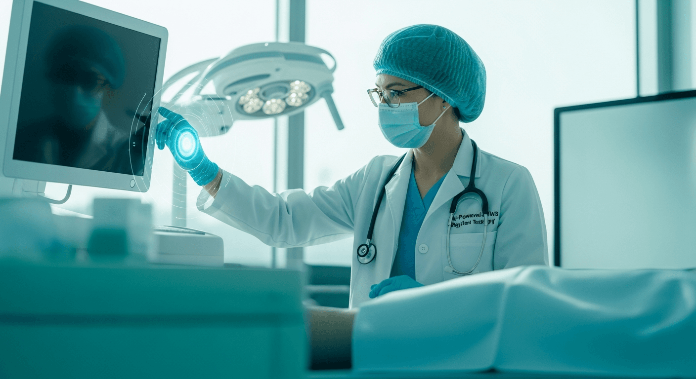
Many believe this new tool could accelerate the discovery of novel therapeutic targets. The ability to test drugs on human-derived retinal cells in a complex, 3D environment may yield more relevant results than traditional models, potentially leading to more effective treatments for conditions like glaucoma, where advances in surgical precision, such as an AI-powered robotic system, are also making strides. While direct clinical applications are still years away, this step is viewed as a crucial foundation for future regenerative medicine strategies.
What Comes Next
While this breakthrough is incredibly promising, it's important to remember that it is currently in the foundational research phase. The next steps will involve further refining the organoid models to ensure they fully mature and accurately mimic all aspects of a human retina. Researchers will also focus on integrating these retinal structures more closely with other neural tissues to study how they connect and transmit visual information to the brain.
Looking ahead, this technology could eventually move towards developing personalized retinal tissue for transplantation. This would involve taking a patient's own cells, growing them into functional retinal tissue in the lab, and then transplanting them into the eye to replace damaged areas. Before reaching clinical trials, however, extensive testing in animal models will be necessary to ensure the safety and effectiveness of such cellular therapies, similar to other complex ophthalmic surgeries like the AI-powered robotic system breakthrough in retinal vein occlusion surgery. This process is typically lengthy, potentially taking several years before human trials can even begin.
When to Talk to Your Doctor
This groundbreaking research represents future potential, not an immediate treatment option. However, if you are experiencing vision changes or managing an existing eye condition, it’s always important to consult your healthcare provider.
Seek immediate medical attention if you experience:
- Sudden vision loss or a dark shadow appearing in your field of vision.
- Flashes of light or new, numerous floaters.
- Eye pain, redness, or sensitivity to light.
If this topic is relevant to a chronic condition you manage, bring this article to your next appointment to discuss whether it changes your care plan.
Sources & Further Reading
Disclaimer: This article is for informational purposes only and does not constitute medical advice. Always consult a qualified healthcare professional.